
Melanoma & Melanoma Surgery in Vienna
Melanoma: Accurate Diagnosis, Complete Surgical Removal, Structured Follow-Up Care
In a nutshell
Malignant melanoma (black skin cancer) originates from the skin’s pigment-producing cells and can spread early on. The thickness of the tumor at the time of removal determines the prognosis.
- Four main types: superficial spreading, nodular, lentigo maligna melanoma, and acrolentiginous.
- Warning signs beyond ABCDE: a mole that looks different from all the others; raised, firm, and growing (EFG); unexplained bleeding.
- Treatment: complete surgical removal, performed on an outpatient basis under local anesthesia; depending on the findings, a second excision with a 0.5 to 2 cm safety margin based on the tumor thickness.
- Follow-up care: frequent during the first three years, then ongoing.
Malignant melanoma —commonly known as black skin cancer —accounts for only a small fraction of all skin cancer cases, but is responsible for most skin cancer-related deaths. The reason lies in its growth pattern: Melanoma can grow deeply and spread at an early stage. Therefore, the thickness of the tumor at the time of removal is crucial. Every millimeter counts.
At my practice in Vienna 1090, I surgically remove suspicious moles under local anesthesia, arrange for a histological examination, and work with you to plan the next steps.
When suspicion arises, time is of the essence
A melanoma cannot be reliably identified with the naked eye. Only a histological examination of the completely removed mole can provide a definitive diagnosis. If melanoma is suspected, I schedule surgery at short notice, discuss each step with you in advance, and plan the incision so that the resulting scar is as inconspicuous as possible.
If you’re just getting started and simply want to have your moles checked: You can find everything you need to know about the process on the page Skin Cancer Screening & Mole Check.
What is melanoma—and why is it so dangerous?
A melanoma develops from melanocytes, the pigment-producing cells of the skin. About two-thirds of all melanomas do not develop from an existing mole, but rather on skin that was previously unremarkable. A mole that has always been there and has not changed is therefore less likely to be a problem than a new spot.
Most melanomas initially grow horizontally, that is, flat within the epidermis. At this stage, they are not yet capable of spreading. Only when the tumor begins to grow vertically into deeper layers does it come into contact with lymphatic and blood vessels.
This is precisely the progression that we want to prevent. The Breslow thickness of the tumor —measured in millimeters from the skin surface to the deepest tumor cell—is therefore the most important single factor for prognosis and determines every subsequent step in treatment.
The Four Most Common Types of Melanoma
Superficial Spreading Melanoma (SSM)
The most common type. It spreads horizontally across the epidermis over a period of months to years before growing deeper—which is why it’s the type of melanoma with the longest window of opportunity for early detection.
Nodular Melanoma (NMM)
It grows downward from the start and forms a lump. About one in twenty melanomas is amelanotic—that is, skin-colored or reddish rather than dark. This type is particularly often detected too late because it does not match the image most people have of skin cancer.
Lentigo Malignant Melanoma (LMM)
It develops on skin that has been damaged by the sun over decades, usually on the face and in older age. It grows very slowly and initially looks like a large, poorly defined age spot.
Acrolentiginous Melanoma (ALM)
It appears on the palms of the hands, the soles of the feet, and under the nails—that is, in areas not exposed to significant sunlight. A new dark vertical streak in the nail that widens or extends onto the cuticle should always be checked out.
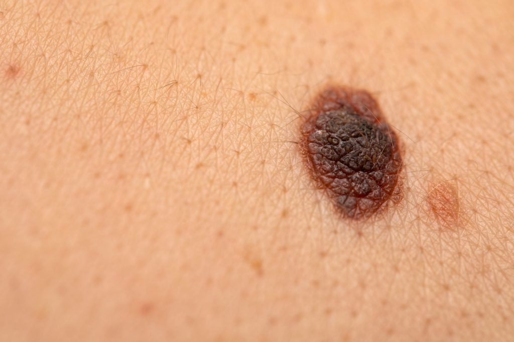
Warning signs that go beyond the ABCDE rule
The ABCDE rule—Asymmetry, Border, Color, Diameter, Evolution—is the best-known mnemonic for self-examination. You can find a detailed explanation of it on my page about mole self-examination.
In practice, however, it doesn’t cover everything. These three warning signs are at least as important:
The Ugly Duckling
A person’s moles usually look similar to one another. If a single mole stands out because it is differently shaped, differently colored, or simply more noticeable than all the others, it should be examined—even if none of the ABCDE criteria apply.
The EFG Rule for Nodular Melanomas
Nodular melanomas are often symmetrical, sharply defined, and uniform in color—the ABCDE rule does not apply here. Instead, the following applies: E—elevated (raised above the skin surface), F—firm (hard to the touch), G—growing (significant increase in size within a few weeks). If all three apply, the lesion should be examined promptly.
Bleeding, itching, and failure to heal
Any spot that bleeds, oozes, or itches for no apparent reason—or that doesn’t heal after a minor injury—is a reason to make an appointment, regardless of how it looks.
From Suspicion to Surgery: The Process
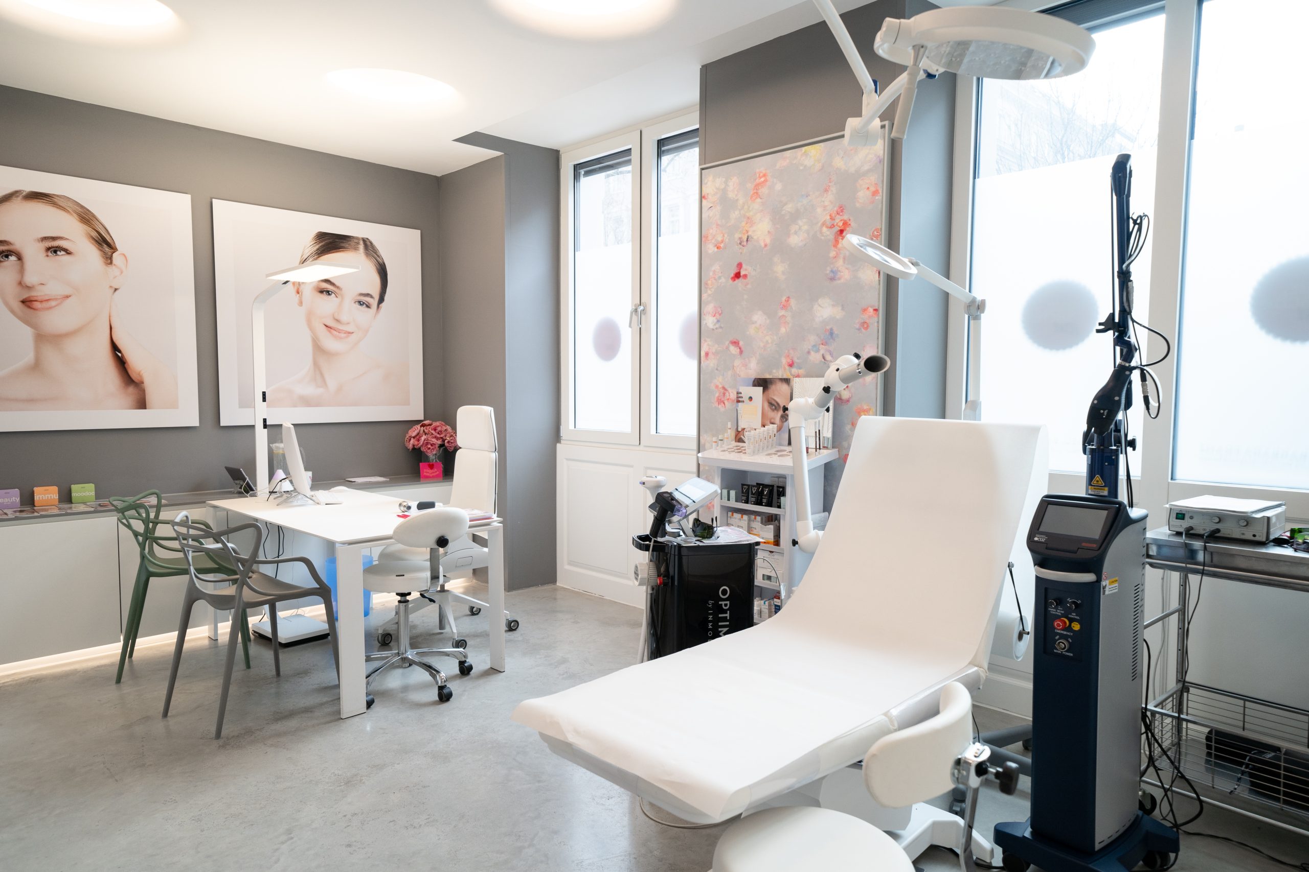
If you come to see me with a suspicious spot, I’ll evaluate it using a dermatoscope and also examine the rest of your skin. I’ve described in detail how a follow-up examination works on the page about mole check .
If the suspicion is confirmed, the lesion will not be incised or excised. In cases of suspected melanoma, complete removal is the standard of care, as this is the only way to reliably determine the tumor’s thickness. You will usually be given a surgery date within a few days.
Melanoma Surgery: What Happens During the Procedure?
Surgical removal is the primary treatment for melanoma. It is performed in two steps—first, the diagnosis; then, the final excision.
1. Excision of a nevus—complete removal
The procedure is performed on an outpatient basis at my office, in compliance with all hygiene guidelines. It is painless for you: I begin by administering a local anesthetic, which you’ll only feel for two to three seconds. I then completely remove the noticeable mark along with a narrow margin of healthy skin. The procedure usually takes 20 to 30 minutes, and you can leave the office immediately afterward.
2. The histological findings
The tissue is sent to a histopathology laboratory. The report not only indicates whether melanoma is present, but also provides the Breslow thickness of the tumor, information on ulceration, and an assessment of the surgical margins. Everything else can be determined from these three pieces of information. As soon as the report is available, I will inform you immediately.
3. Secondary excision with a safety margin
If the test results confirm melanoma or one of its precursor lesions, a second procedure is performed. During this procedure, the area is excised again with a defined safety margin to ensure that no abnormal cells remain in the surrounding tissue. The size of this margin depends on the test results:
- Precursor (dysplastic nevus): complete removal with a narrow margin; further treatment is usually not necessary
- Melanoma in situ (confined to the epidermis): 0.5 cm
- Tumor thickness up to 2 mm: 1 cm
- Tumor thickness greater than 2 mm: 2 cm
The safety margin is measured in width, not in depth—so the wound becomes wider but not deeper.
This illustrates why early detection is so crucial: The earlier a lesion is removed, the less invasive the procedure will be and the better the prognosis. For precancerous lesions and melanomas that are still confined to the epidermis, treatment is generally considered complete once the lesion has been completely removed.
4. Sentinel Lymph Node (Sentinel Biopsy)
Once the tumor reaches a certain thickness, the first lymph node in the drainage area is also examined to determine whether cells have already spread. This procedure is performed in a specialized department—I will arrange the referral and guide you through the rest of the process.
5. Wound Closure and Scar Formation
The scar is always longer than the removed area because the edges of the wound must come together without tension. I make the incision along the natural lines of skin tension so that the scar later disappears into an existing skin fold rather than running across it.
For larger defects, direct closure is not sufficient. In such cases, flap procedures —involving the repositioning of adjacent tissue—or a skin graft are used. The appropriate technique depends on the location and size of the defect and will be discussed with you before the procedure.
All of this applies to confirmed melanomas, where playing it safe is the top priority. For all other skin lesions, I take a different approach—more on that in the next section.
Wound healing without stitches—a focus of my practice
Whenever medically appropriate, I treat wounds without sutures. Current guidelines recommend that many skin lesions do not require sutures. The wound then heals on its own from the bottom up, leaving virtually no scar in many cases.
The reason is simple: Every incision that is sutured leaves a scar that remains permanently. A wound that heals openly and in a controlled manner does not.
Whether this technique is appropriate depends on the location, size, and type of skin lesion. In the case of a confirmed melanoma, complete removal with a safety margin takes priority; in such cases, neat wound closure is necessary. In contrast, for inconspicuous and benign moles, it is very often possible to perform the procedure without sutures.
After Surgery: Wound Care and Follow-Up Care
For the first few days, keep the wound dry and take it easy. Avoid exercise, saunas, swimming pools, and intense sun exposure until the stitches are removed. I will discuss the specific guidelines with you individually.
After melanoma treatment, structured follow-up care is important. It involves examining the entire skin, the scar, and the lymph node regions. The frequency of these checkups depends on the stage of the disease and is most frequent during the first three years, as most recurrences occur during this period.
People who have already had a melanoma also have a significantly increased risk of developing a second, unrelated melanoma. Regular checkups therefore remain an ongoing part of treatment.
It takes a year for a fresh scar to reach its final appearance. If it thickens or remains bothersome during this time, there are treatment options—learn more on my page about scar treatment.
Who is at increased risk for melanoma?
- Fair skin, reddish or blond hair, freckles
- More than 50 moles or unusual, atypical moles
- A history of melanoma in oneself or in one's parents, siblings, or children
- Severe sunburns, especially during childhood and adolescence
- Regular use of tanning beds, especially before the age of 35
- A weakened immune system, such as after an organ transplant
If any of these points apply to you, I recommend getting checked more often than once a year —we can discuss the appropriate frequency together. I’ve summarized what else you can do on your own to protect your skin under “Skin Cancer Prevention.”
Frequently Asked Questions About Melanoma
Is melanoma curable?
Yes. If a melanoma is detected early and completely removed surgically, the prognosis is very good—for thin melanomas confined to the epidermis, surgery usually cures the disease. The key factor is the thickness of the tumor at the time of removal.
How fast does a melanoma grow?
It depends on the type. A superficial spreading melanoma initially grows flat over a period of months to years. A nodular melanoma, on the other hand, can grow deep into the skin within a few months. As a general rule, any change you notice within a few weeks should be evaluated promptly.
Does melanoma hurt?
Usually not. That’s exactly what makes it dangerous—melanoma usually causes no symptoms. Some melanomas itch, bleed slightly, or don’t heal after minor injuries. Don’t rely on pain as a warning sign; instead, look for changes in appearance.
How long does the surgery take?
The removal of a conspicuous birthmark usually takes 20 to 30 minutes and is performed on an outpatient basis under local anesthesia. You can go home immediately afterward. A subsequent excision with a safety margin takes a little longer, but follows the same procedure.
Is the procedure painful?
The area will be numbed with a local anesthetic; you’ll only feel the brief prick of the needle. You won’t feel any pain during the surgery. In the first one to two days afterward, the area may feel slightly tight or tingly—usually a simple pain reliever is enough, and often none is needed at all.
Why is a second surgery necessary?
During the first surgery, the lesion is removed with a narrow margin because the required size of the safety margin can only be determined from the histological findings. Only once the tumor thickness is known can the final margin be determined. The second procedure is therefore not an indication that anything was overlooked during the first one.
Do I need to go to the hospital for the melanoma surgery?
In most cases, no. Removal and re-excision are performed on an outpatient basis at my office in Vienna 1090. Inpatient surgery is necessary only if a larger defect needs to be closed or if a sentinel lymph node biopsy is scheduled.
Can melanoma develop even without exposure to the sun?
Yes. Melanomas can also occur on the soles of the feet, under the nails, on the scalp, or on mucous membranes—in other words, in areas that are practically never exposed to the sun. UV radiation is the most important risk factor, but it is not the only one. Genetic predisposition also plays a role.
Does health insurance cover the costs?
I am a private practice physician and do not have contracts with health insurance providers. After your treatment, you will receive an invoice that you can submit to your health insurance provider. A portion of the costs is usually reimbursed. The exact amount depends on your insurance provider and the services rendered.

